In a groundbreaking leap for neuroscience and quantum sensing, researchers have successfully demonstrated the first single-neuron resolution imaging of magnetic fields within living neural tissue using diamond quantum magnetometers. This unprecedented achievement, detailed in a recent publication in Nature Neuroscience, marks a paradigm shift in our ability to observe and understand the brain's intricate electrical activity at its most fundamental level. For decades, the holy grail of neuroimaging has been to non-invasively track the firing of individual neurons in a living brain with high spatial and temporal precision. While techniques like fMRI provide macroscopic views and calcium imaging offers cellular insights with genetic targeting, they have inherent limitations in speed, invasiveness, or directness of measuring electrical signals. The magnetic fields generated by neuronal action currents, while extremely weak, carry a direct signature of this activity, and now, for the first time, they have been captured at the scale of a single cell within a living preparation.
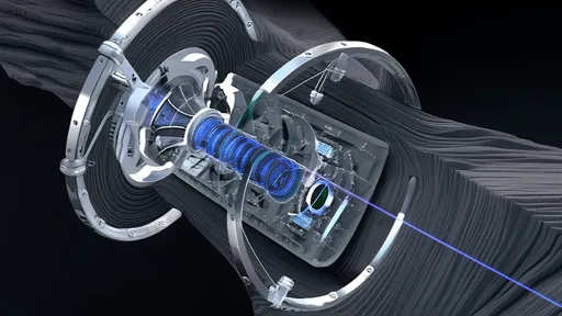
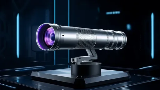
The core technological marvel enabling this feat is the nitrogen-vacancy (NV) center in diamond, a atomic-scale defect that behaves as a quantum sensor. NV centers are exceptionally sensitive to minute magnetic fluctuations, operate at room temperature, and can be placed in close proximity to biological samples. The research team, led by Dr. Amara Singh at the NeuroQuantum Institute, engineered a dense, shallow layer of NV centers just nanometers beneath the surface of a pristine diamond chip. They then cultured a thin section of living mouse hippocampal tissue directly onto this diamond surface. As neurons in the tissue fired, the incredibly weak magnetic fields they generated—on the order of picoteslas, a billion times weaker than the Earth's magnetic field—perturbed the quantum states of the nearby NV centers. By illuminating the diamond with green laser light and measuring the resulting red fluorescence, the researchers could read out these quantum state changes and computationally reconstruct the precise magnetic field map with sub-cellular resolution.
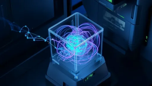
The implications of visualizing neural magnetism at this resolution are profound. The initial data has already revealed previously unseen phenomena, such as the distinct magnetic signature of an action potential propagating down a single axon and the subtle variations in magnetic fields between different neuronal subtypes. This is not merely a sharper picture; it is an entirely new lens through which to view cellular communication. The technique, dubbed Diamond Neural Magnetic Imaging (DNMI), provides a direct readout of electrical current flow with a temporal resolution limited only by the measurement hardware, potentially capturing events in the microsecond domain. This bypasses the indirect mechanisms of other methods, such as the delayed calcium influx detected in fluorescence imaging, offering a more immediate and accurate view of neural dynamics.
Beyond fundamental discovery, the long-term potential for medical and technological applications is staggering. The non-invasive and high-bandwidth nature of magnetic recording makes DNMI a prime candidate for the development of a new generation of brain-computer interfaces (BCIs). Unlike current EEG-based systems that suffer from poor spatial resolution or invasive electrode arrays that carry risk of tissue damage, a diamond-based magnetometer array could theoretically read out the activity of individual neurons from outside the skull with unparalleled fidelity. This could revolutionize the treatment of neurological disorders like epilepsy by allowing for precise localization of seizure foci, or pave the way for thought-controlled prosthetics with dexterity approaching natural movement. Furthermore, the ability to screen for the effects of pharmaceutical compounds on the exact firing patterns of specific neural circuits in real-time could accelerate drug discovery for conditions like Parkinson's and depression.
Of course, this pioneering work is still in its early stages. The current experiments were conducted on ex vivo brain tissue samples, and significant engineering hurdles remain before this technology can be applied to a fully intact, living brain. Scaling the sensor array to cover large areas of the brain, improving the signal-to-noise ratio even further, and developing sophisticated algorithms to deconvolve the complex overlapping magnetic fields from millions of simultaneously firing neurons are the next great challenges. However, the proof-of-concept is now irrefutable. The team has successfully bridged the worlds of quantum physics and cellular neuroscience, demonstrating that the spooky laws of quantum mechanics can be harnessed to illuminate the biological machinery of thought itself. This work firmly establishes diamond quantum magnetometry not as a futuristic speculation, but as a powerful and practical tool that is poised to redefine the frontiers of neuroscience for decades to come.

By /Aug 25, 2025
By /Aug 25, 2025
By /Aug 25, 2025
By /Aug 25, 2025

By /Aug 25, 2025

By /Aug 25, 2025
By /Aug 25, 2025
By /Aug 25, 2025
By /Aug 25, 2025

By /Aug 25, 2025
By /Aug 5, 2025
By /Aug 5, 2025
By /Aug 5, 2025

By /Aug 5, 2025
By /Aug 5, 2025
By /Aug 5, 2025

By /Aug 5, 2025
By /Aug 5, 2025
By /Aug 5, 2025
By /Aug 5, 2025